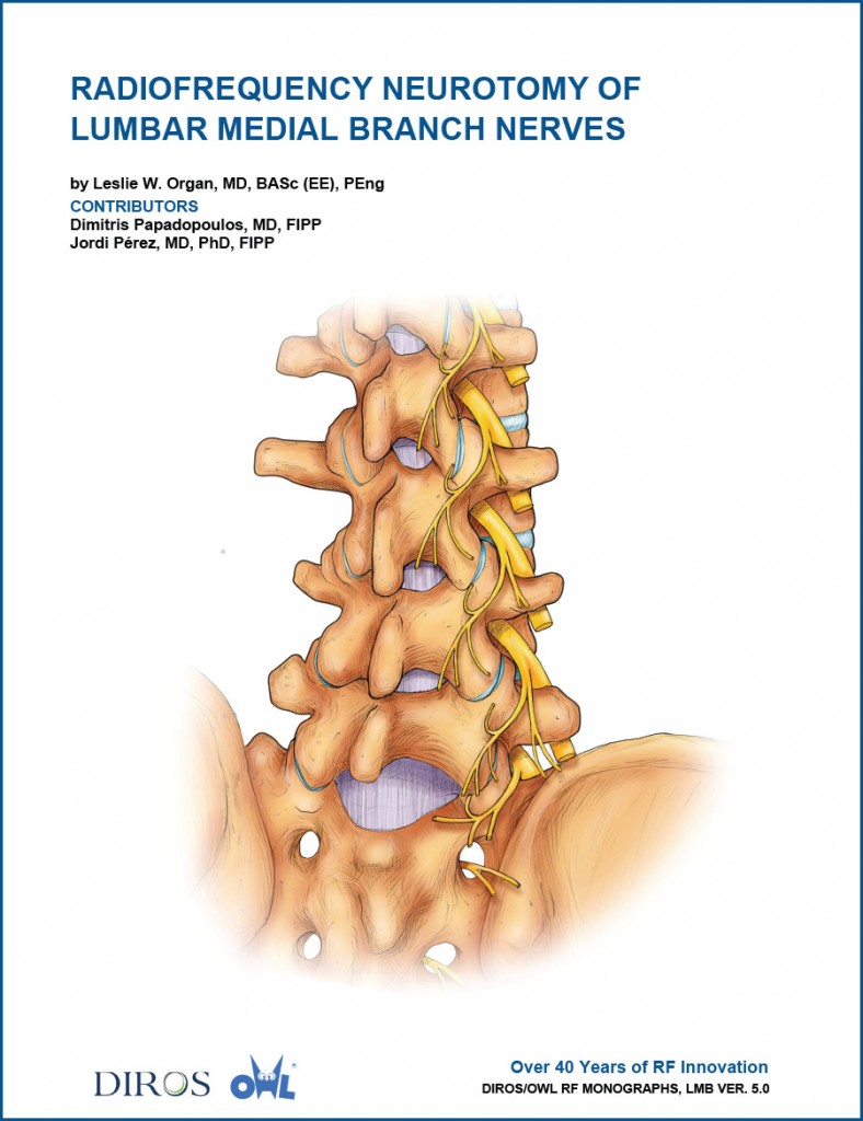
Radiofrequency Neurotomy of
Lumbar Medial Branch Nerves
by Leslie W. Organ, MD, BASc (EE), PEng, Dimitris Papadopoulos, MD, FIPP, and Jordi Pérez, MD, PhD, FIPP
This 26 page procedural monograph clearly describes, with the use of original full color drawings, the anatomy and neuroanatomy of the lumbar facet joints and L5/S1 joint. It also provides practical information such as facilities and equipment required and guidelines for patient preparation, positioning, and C-arm fluoroscopy use. Vertebral and radiological landmarks for precise electrode positioning are detailed and illustrated, and the importance of special structures such as the mamillo-accessory ligament relative to successful facet joint RF denervation are identified.
Monograph Contents, include:
- Anatomy of the facet joints and the medial branch nerve
- Diagnosis
- Patient history and examination
- Diagnostic blocks
- Facilities and equipment
- Guidelines for patient preparation
- Positioning of the patient and the C-arm fluoroscopy unit
- Radiology of the lumbar medial branch
- Vertebral landmarks
- Radiological landmarks
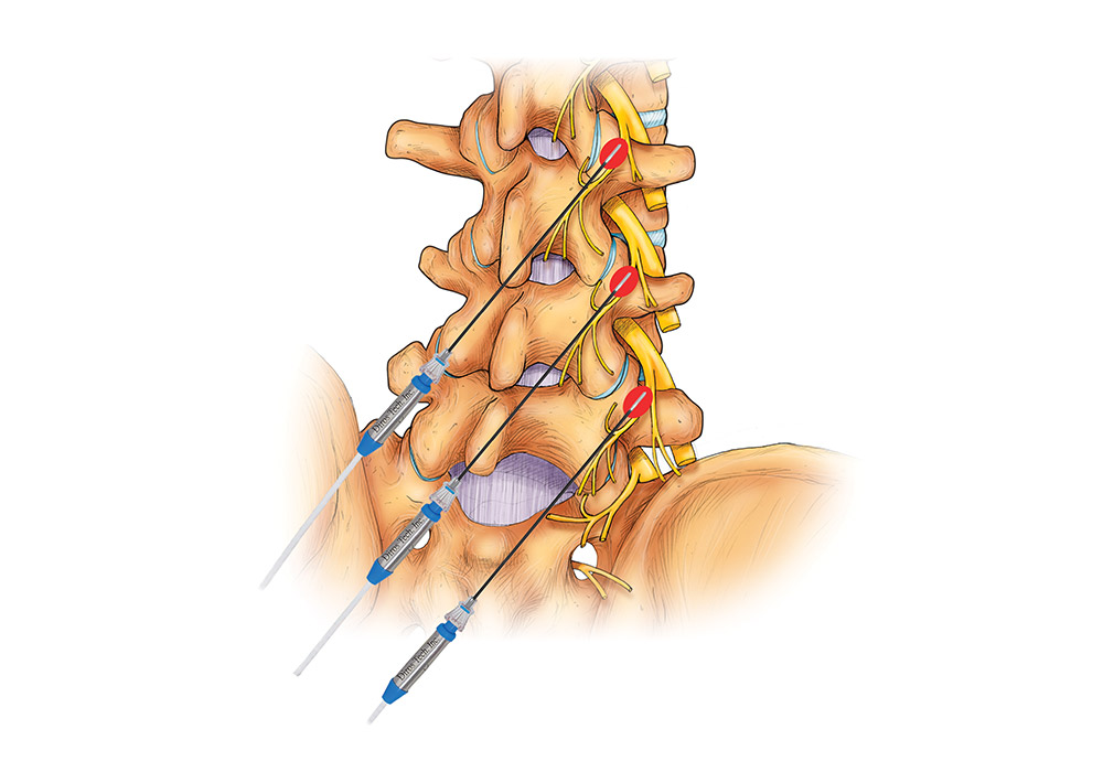
- Denervation of lumbar facet joints
- Approach to the medial branch nerve
- The mamillo-accessory ligament
- Electrical stimulation
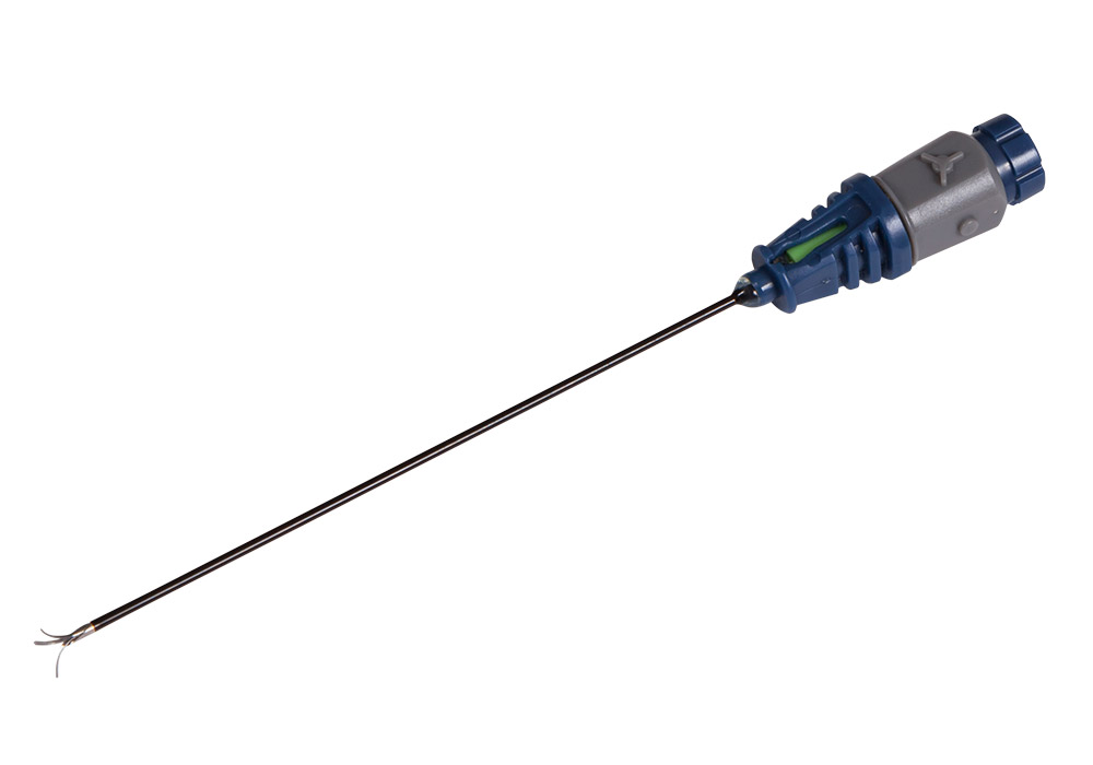
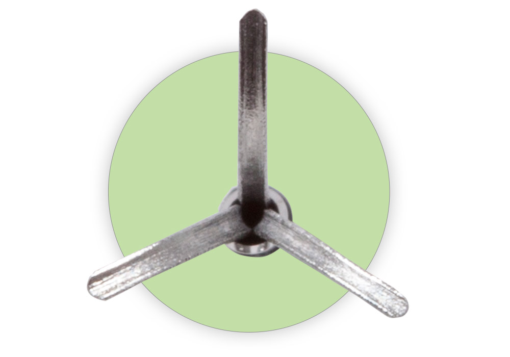
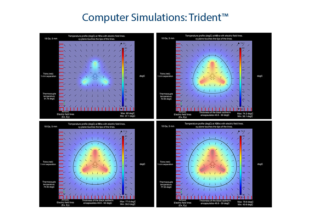
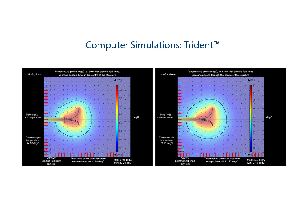
- RF thermal ablation
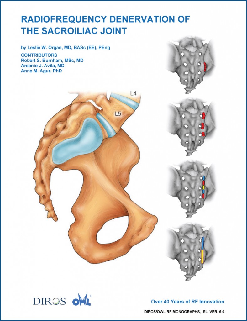
Radiofrequency Denervation of the Sacroiliac Joint
by Leslie W. Organ, MD, BASc (EE), PEng, Robert S. Burnham, MSc, MD, FRCPC, Arsenio J. Avila, MD, FRCP, FCCP, and Anne M. Agur, PhD
This 41 page procedural monograph clearly describes, with the use of original, high quality full color drawings, the anatomy and unique neuroanatomy of the posterior sacroiliac joint and associated ligaments. It features tables that provide not generally available information such as posterior sacral foraminal height and interforaminal distance to allow calculation of S1 to S3 distance as required for linear strip RF lesions of lateral branch (LB) nerves of the posterior sacral rami, and another table presenting detailed information of the numbers and directions of exiting LB nerves from the foramina of L5/S1 and S1 to S4.
Monograph Contents, include:
- Anatomy and innervation of the sacroiliac joint
- Diagnosis
- Patient history and examination
- Manual Provocative tests
- Diagnostic anesthetic blocks
- Facilities and equipment
- Guidelines for patient preparation
- Target identification
- Fluoroscopy
- Ultrasonography
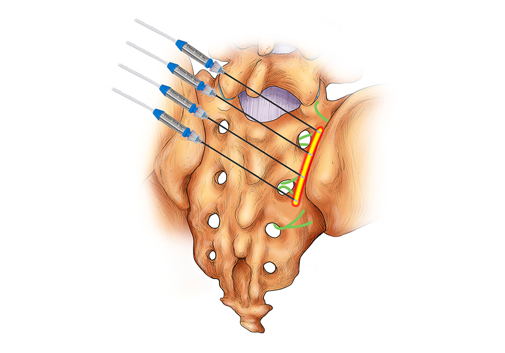
- Techniques of RF denervation of the sacroiliac joint:
- Cooled RF periforaminal
- Conventional RF periforaminal
- Bipolar linear strip lesion
- Tripolar linear strip lesion
- Quadrapolar linear strip lesion